Saturday, September 4, 2010

NECROTIZING ENTEROCOLITIS


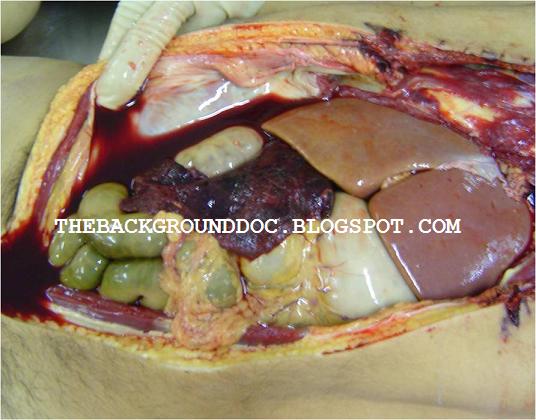
This male neonate died after 48 hours after delivery with abdominal distention and fever.
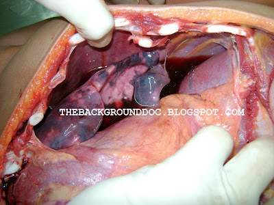
There was no abnormal history during pregnancy and delivery. A wrong clinical diagnosis of hepatosplenomegaly was made. A full autopsy was performed. On external examination, significant abdominal distention was noted. Internal examination revealed pneumoperitoneum and large bowel necrosis with multiple submucosal gas-filled cysts. The other organs were grossly normal.

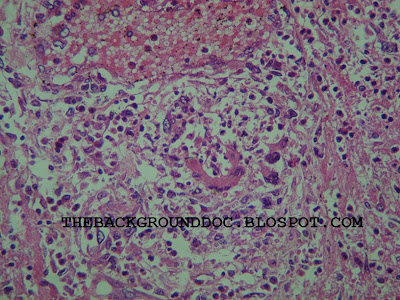
Pneumatosis intestinalis when present, is diagnostic of necrotizing enterocolitis. It is thought to be due to the production of gas from bacterial fermentation of substrate with a significant portion being hydrogen gas.

Necrotizing enterocolitis (NEC) occurs primarily in premature low birth-weight infants who have undergone some form of perinatal stress.

ANTAL GENERSICH

Antal Genersich (1842-1918), was a hungarian pathologist who first described a case with signs and symptoms similar to NEC in a 45 hour old premature infant who died within 24 hours (Genersich A: Bauchfellentztindung beim Neugeborenen in. Folge yon Perforation des Ileums. Arch Pathol Anat 120:485, 1891.)

He was the founder of the Anatomical Pathology Institute in Cluj/Kolozsvar. A disciple of Rokitansky and Virchow, Genersich was a great theorist but also a clinical doctor.


Thursday, August 19, 2010

PURPURA FULMINANS

Necropsy performed in a female infant with a generalized purpuric rash. Internal examination revealed dark red adrenal glands without changes in their size and position. Other organs were slightly congested. There is no evidence of meningitis. This patient exhibited the sudden onset of the Waterhouse-Friderichsen syndrome: cyanotic pallor, petechial rash and...death.

Waterhouse-Friderichsen Syndrome
Waterhouse-Friderichsen syndrome is an acute adrenal insufficiency due to massive haemorrhage into the adrenal gland, more often bilateral, caused by malignant form of meningitis. It occurs in fulminant bacterial infections, due principally to the meningococcus, influenza or colon bacillus.


First described in 1894 by Arthur Francis Voelcker (1861-1946; left) and in 1901 by the British dermatologist Ernest Gordon Graham Little (1867-1950; right).


It was first reported as an entity by Rupert Waterhouse (photo) in 1911, and the subject was comprehensively reviewed in 1918 by the Danish paediatrician Carl Friderichsen.


Saturday, August 14, 2010

BLUE DISEASE

Here, an example of Tetralogy of Fallot with pulmonary atresia (pseudotruncus arteriosus), a severe variant in which there is complete obstruction (atresia) of the right ventricular outflow tract, causing an absence of the pulmonary trunk during embryonic development. We see a complex heart malformation in a three-month boy: overriding aorta and ventricular septal defect (VSD).In these individuals, blood shunts completely from the right ventricle to the left where it is pumped only through the aorta. The lungs are perfused via extensive collaterals from the systemic arteries, and sometimes also via the ductus arteriosus.
Photo: www.fotosearch.co.kr/LIF154/ccp06018
Text: http://en.wikipedia.org/wiki/Tetralogy_of_Fallot


ÉTIENNE-LOUIS ARTHUR FALLOT

Étienne-Louis Arthur Fallot (1850-1911) was a French physician born in Sète. Etienne Fallot attended medical school in Montpellier in 1867. While in residence in Marseille he wrote a thesis on pneumothorax. In 1888 he was made Professor of Hygiene and Legal Medicine in Marseille. In 1888 Etienne Fallot accurately described in detail the four anatomical characteristics of tetralogy of Fallot.
SOURCE: http://en.wikipedia.org/wiki/Etienne_Fallot

TEN PERCENT TUMOR

A 37-year-old female died after an episode of emesis, headache, chest pain and high arterial pressure. A diagnosis of myocardial infarction without ST-segment elevation was made. Despite the use of antihypertensive therapy, blood pressure remained high. She died after losing consciousness. Autopsy showed a ruptured left adrenal tumor and a morphologically normal heart. Tumor was well encapsulated and formed by zellballen nests of polygonal shaped cells with finely granular cytoplasm. Peritumoral brown fat reflects adrenergic stimulation. This is a case of adrenergic crisis due to spontaneous rupture of a sporadic pheochromocytoma.

LUDWIG PICK

"The first description of the adrenal gland dates since 1563 by Eustachius in a study "Opuscula anatomica". Despite earlier recognition of the presence of adrenals and division into cortex and medulla, the precise observations of Addison appeared in 1855 that the essential role of these glands was recognized in patients who died with adrenal destruction secondary to tuberculosis. Frankel first described a medulary adrenal tumour in 1886. In 1912 the pathologist Pick formulated the descriptive term pheochromocytoma. The first surgical removal of pheochromocytoma in Europe was performed by Rouks in 1926 and by Mayo in USA in 1927."
Srp Arh Celok Lek. 2002 Jul;130 Suppl 2:31-7.

Professor Ludwig Pick (August 31, 1868 - February 3, 1944) was a German pathologist who was a native of Landsberg an der Warthe. In 1893 he earned his medical doctorate in Leipzig, and subsequently practiced medicine at Leopold Landau's private Frauenklinik, where he remained until 1906. That same year he became director of the department of pathological anatomy at the city hospital Friedrichshain-Berlin. Later, he was imprisoned by the Nazis, and died on February 3, 1944 at the Theresienstadt Concentration Camp.
Ludwig Pick made several contributions to academic pathology, particularly in the field of genitourinary diseases, and also in the study of melanotic pigmentation. In 1912 he coined the term pheochromocytoma to describe the chromaffin color change in tumor cells associated with adrenal medullary tumors.

Saturday, April 10, 2010

Thrombotic Thrombocytopenic Purpura


A fatal case of thrombotic thrombocytopenic purpura (TTP) is described here. A 27-year-old man presented with thrombocytopenia, facial purpura, arthritis and daily fever for two months. A week before death, he was admitted to a hospital and treated, including plasma infusion and plasma exchange. However, he died of pneumonia, evidence of cardiac tamponade and pancreatitis.
At autopsy, there were mild jaundice, cerebral edema, mediastinal hematoma, hydrothorax and pulmonary condensation, splenic infarction, hemoperitoneum and hemorrhagic pancreatitis. Microscopically, disseminated microvascular hyaline platelet thrombi were prevalent in terminal arterioles and capillaries of the heart and kidneys, brain, spleen (splenic infarction) and pancreas (pancreatitis). In several organs, megakaryocytes were distributed (increased thrombocytopoiesis).


MOSCHCOWITZ'S DISEASE
Thrombotic thrombocytopenic purpura (TTP) is also known as Moschcowitz's Disease.

ABOUT THE EPONYM: ELI MOSCHCOWITZ, M.D.
Dr. Eli Moschcowitz, of New York City, died on February 23, 1964, at the age of eighty-two. Dr. Moschcowitz graduated from Columbia University College of Physicians and Surgeons in 1900. He was a consulting physician at Beth-El and the Mount Sinai Hospitals and a consulting pathologist at Beth Israel Hospital. In 1925, he described a vascular disease involving blood clots which is named Moschcowitz's Disease after him (An acute febrile pleiochromic anemia with hyaline thrombosis of terminal arterioles and capillaries: An undescribed disease.Archives of Internal Medicine, Chicago, 1925, 36: 89). In 1945, Dr. Moschcowitz retired as Director of the Mount Sinai Hospital and as Professor of Clinical Medicine at Columbia University College of Physicians and Surgeons. He was an Associate Member of the Medical Library Association, a Diplomate of the American Board of Internal Medicine, and a member of numerous societies, including the New York Academy of Medicine and the American Association of Pathologists
and Bacteriologists. Reference: Bull Med Libr Assoc. 1965 April; 53(2): 302. (http://www.ncbi.nlm.nih.gov/pmc/articles/PMC198274/).
Photo: http://www.ttpnetwork.org.uk/WhatIsTTP.asp

Sunday, May 31, 2009

WEGENER'S GRANULOMATOSIS

A 38-YEAR-OLD WOMAN WITH ARTHRITIS, PURPURIC RASH AND PULMONARY HEMORRHAGE.
Over a period of six months, this patient presented with symptoms related to arthritis, hematuria, oral ulceration, purpuric rash and occasional hematemesis. She remained untreated.
Some hours before death, she was hospitalized in a tertiary hospital. At that moment, serum creatinine level was found to be increased (6,9mg/dL). While on dialysis, she died after voluminous discharge of blood from the orotracheal tube.

At necropsy, purpuric rash on the upper and lower extremities was evident. Other findings included oral ulcerations, massive pulmonary hemorrhage (1050g, left lung; 1200g, right lung), pale kidneys and confluent serpiginous areas in the spleen (220g).
Under microscope, alveolar capillaritis and focal segmental necrotizing glomerulonephritis with crescent formation were observed. Necrosis and granulomatous vasculitis occurred in the spleen and palate. The purpuric lesions correspond to leukocytoclastic vasculitis.


SEE VIDEO: http://www.youtube.com/watch?v=-sNcAiRWLTU&feature=related


ABOUT THE EPONYM: FRIEDRICH WEGENER
From Wikipedia, the free encyclopedia
Friedrich Wegener (1907-1990) was a German pathologist who is notable for his description of a rare disease. Although this disease was known before Wegener's description, since the 1950s it has been called by the name Wegener’s granulomatosis.
Wegener joined the Nazi Party in 1932. As a relatively high ranking military doctor he spent some of the war in a medical office three blocks from the Lodz Ghetto, a Jewish ghetto in Lodz, Poland. There is speculation that he participated in experiments on concentration camp inmates.The American College of Chest Physicians (ACCP) awarded Wegener a “master clinician” prize in 1989. After his Nazi past was discovered in 2000, the ACCP rescinded the prize and, separately, a campaign was begun to rename Wegener’s granulomatosis to ANCA-associated granulomatous vasculitis.
READ MORE:

HEINRICH RUDOLF HERTZ AND WEGENER'S GRANULOMATOSIS
The hertz standard unit was named in honor of Heinrich R. Hertz, a German physicist who was a pioneer in the study of electromagnetic radiation.
Heinrich Hertz is credited with the discovery of electromagnetic waves. Hertz performed numerous experiments and provided a wealth of data concerning reflection, refraction, and interference with regards to electric waves.
Heinrich Hertz died in 1892 of Wegener's granulomatosis at the age of 36.

Monday, May 11, 2009

FORGET SWINE FLU

DENGUE FEVER OUTBREAK IN NORTHEAST BRAZIL KILLS 47


RIO DE JANEIRO, April 28 (RIA Novosti) - An ongoing dengue fever outbreak in northeast Brazil has claimed 47 lives since January, the country's Health Ministry said on Monday.
According to ministry statistics, 55,000 people in Bahia, the fourth most populous Brazilian state, are currently registered as dengue fever sufferers, a 220% increase from the same period of last year.
Authorities have declared a state of emergency in seven cities and requested aid from the federal government to cope with the disease.
Dengue is passed from human to human via mosquitoes or blood transfusions. Its symptoms are the sudden onset of severe headache, high fever, and muscle and joint pains, and are often diagnosed as influenza. It is also known as break-bone fever or bonecrusher disease due to severe pain experienced in the joints.
According to the World Health Organization, the disease is now epidemic in more than 100 countries.

FORGET SWINE FLU

A dengue-seropositive child with submassive pulmonary hemorrhage and serohemorrhagic pleural effusion.

Copyright (mosquito): ABC TV News

Wednesday, April 22, 2009

SU(nday)DDEN DEATH


THIS ENLARGED HEART DOES NOT BEAT ANYMORE IN THE CHEST OF A YOUNG MAN.
The images reveal a hypertrophic and dilated heart weighing 1100g: a cardiomyopathy supposed to be idiopathic. Note the nutmeg liver (3000 grams) expressing the end-stage congestive heart failure. He died last Sunday at 33 years of age.
To understand the cardiac liver: http://www.youtube.com/watch?v=a2L6gwoOw70
The nutmeg liver:
The real nutmeg:

Saturday, April 11, 2009

A BLEEDING GASTRIC ULCER


The images show a deep stomach ulcer that bled causing sudden and profuse hematemesis. Stomach and bowel were distended by bloody content. The cause of death was hypovolemic shock.

Saturday, April 4, 2009

HEREDITARY HEMOCHROMATOSIS

An elderly diabetic man died after an episode of abdominal pain and fever that lasted two days. He was known to have a “disease in the liver” and biliary lithiasis. In fact, he had liver cirrhosis as seen at autopsy. There were also dark gallstones and a brown-pigmented pancreas. The cause of death was acute pancreatitis. Microphotograph shows excessive amounts of iron stores within hepatocytes (Perls prussian blue stain).


Perls stain is the oldest histochemical technique still in use (Max Perls, 1846).